Introducing the Art + Science Pathway

By Sophie Lynford, Kate Smith
May 27, 2022
Index Magazine

Introducing the Art + Science Pathway

Two side-by-side images each show a sculpture of a falcon wearing a crown. The sculpture is atop a small platform. The images face each other. The image at left is a black and white X-radiograph, which reveals internal elements of the sculpture. The image at right is a color photograph and shows the sculpture as brownish-green set against a neutral gray ground.
An X-radiograph of an Egyptian bronze falcon sculpture (dating to the mid-7th to late 6th century BCE) revealed hidden features to conservators in the museums’ Straus Center for Conservation and Technical Studies.

On your next visit to the museums, keep an eye out for our new Art + Science Pathway in the permanent collections galleries. The pathway highlights objects that have been treated and studied by conservators and conservation scientists at the Harvard Art Museums.

Artistic and scientific inquiry have always been deeply intertwined at the museums. Each year, more than 8,000 objects are conserved and analyzed in the Straus Center for Conservation and Technical Studies. The first facility of its kind in the United States, the Straus Center houses labs that specialize in the conservation of works on paper, paintings, sculpture, decorative objects, and historical and archaeological artifacts. 

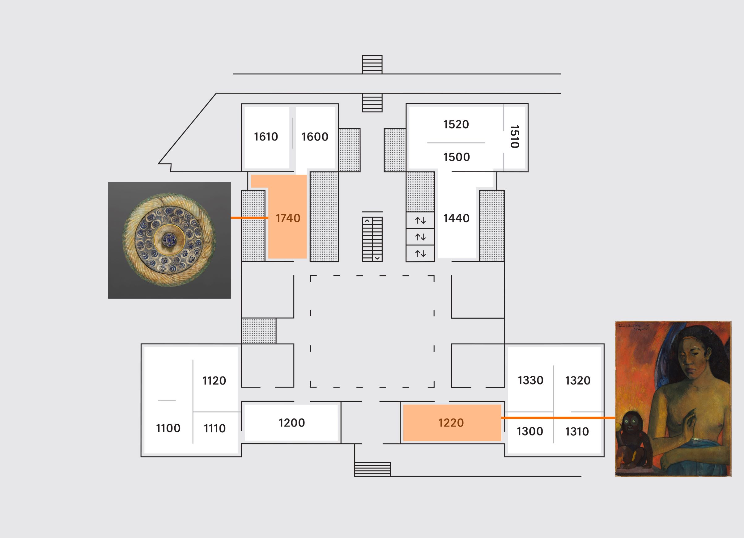
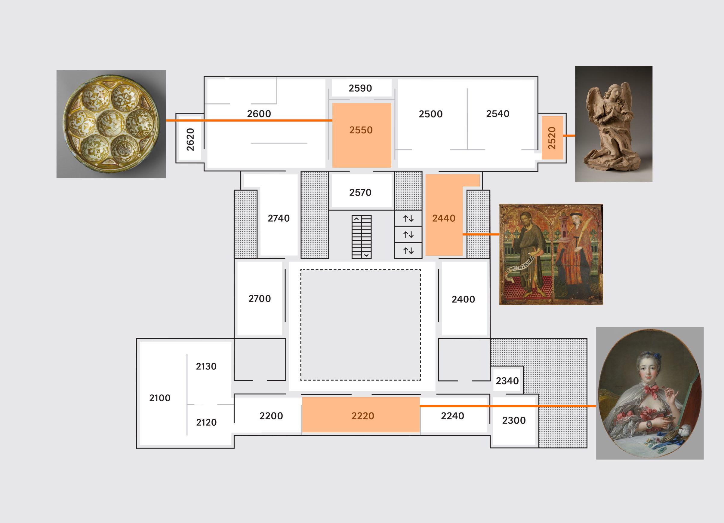
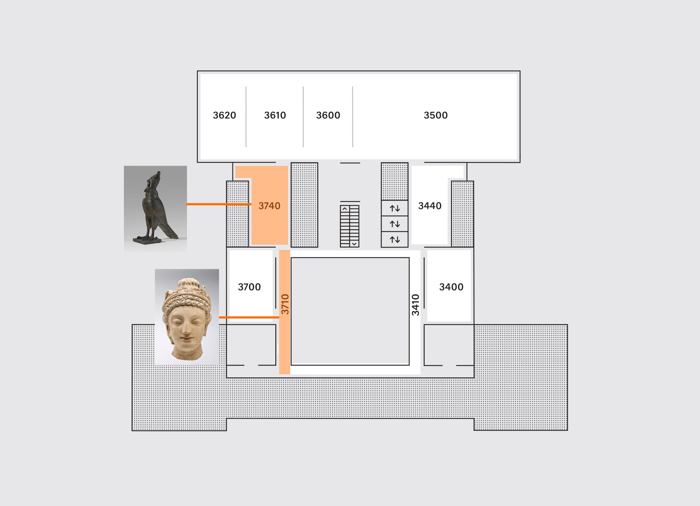
Technical imaging and scientific analysis help resolve long unanswered questions, yield insights, and uncover hidden stories about artworks. We invite you into the galleries to explore the pathway—highlighted in the maps below and called out in the galleries with orange brackets around the wall labels—to learn how objects were made, how they have changed over time, and what has been discovered beneath the surface. 

The first group of objects on the Art + Science Pathway was examined through radiography, or X-rays. This imaging technique tells us about inner structures of objects, just as it does with the human body. The X-rays penetrate light materials and are absorbed by dense materials, producing maps of relative density. A radiograph can make visible parts of an object that an artist painted over, because both the underpainting and the surface layer block radiation. It can also provide details about the surface on which an artist painted, such as the weave of a canvas or the grain of a wood panel. X-radiographs of three-dimensional objects will show old breaks that have been covered up by restoration, or the way a sculptor used clay or stone to build around a metal support structure. Across our galleries, eight objects are accompanied by reproductions of radiographs as well as explanations of what the X-rays revealed about the works.

Pathway objects can be found in the following galleries:

Level 1

Gallery 1220
Paul Gauguin, French, Poèmes Barbares, 1896. An X-radiograph of this painting reveals a different image below the surface.

Gallery 1740
Mirror, China, 20th-century pastiche using 3rd–2nd century BCE components. An X-radiograph shows a highly decorated cast surface of a bronze mirror hidden beneath ornamentation in precious materials.

Level 2

Gallery 2220
François Boucher, French, Pompadour at Her Toilette, 1750, with later additions. In its early decades, this painting was reshaped multiple times. These modifications are recorded in the X-radiograph of the work. 

Gallery 2440
Lluís Borrassà, Spanish, Saint John the Baptist and Saint Barbara, c. 1411–13. This panel painting’s X-radiograph reveals elements of its construction, including wooden battens that hold planks together and coarse fabric glued over the joins. 

Gallery 2520
Gian Lorenzo Bernini, Italian, Kneeling Angel, 1672. The X-radiograph of this sculpture indicates that the artist modeled it principally from one piece of solid clay. 

Gallery 2550
Sweetmeat dish, Iran, Seljuk-Atabeg period, c. 1200. A computed tomography scan, which captures multiple X-radiographs from different views, shows the extent to which this dish had been reconstructed over the years.

Level 3

Gallery 3710
Head of a bodhisattva, Afghanistan, Hadda, Gandharan, 4th–5th century. A micro-computed tomography scan shows the mixture of coarse minerals that comprises the bulk of the head’s solid form. 

Gallery 3740 
Horus falcon wearing crown of Upper and Lower Egypt with uraeus, Egypt, Late Period, Dynasty 26, mid-7th to late 6th century BCE. Radiography uncovered small bones, likely from a bird, encased in this bronze falcon. 

Two side-by-side images each show a sculpture of a falcon wearing a crown. The sculpture is atop a small platform. The images face each other. The image at left is a black and white X-radiograph, which reveals internal elements of the sculpture. The image at right is a color photograph and shows the sculpture as brownish-green set against a neutral gray ground.
Art Talk—Art + Science Pathway with Kate Smith

In the coming months, we’ll add a group of objects to the pathway to show the impact of conservation treatment. Alongside recently conserved objects, you’ll find photographs of the works before they were treated to illustrate the ways deterioration can be stabilized or reversed.


Sophie Lynford is the Rousseau Curatorial Fellow in European Art, and Kate Smith is conservator of paintings and head of the paintings lab in the Straus Center for Conservation and Technical Studies at the Harvard Art Museums.